
Differentiating AV Blocks
Objective: Use a simple three-question path to tell 1°, Mobitz I, Mobitz II, and complete heart block apart — with a CardioStrip example for each.
Why This Matters
AV blocks all look like “something is wrong between the P and the QRS.” Under pressure, that makes them easy to mix up. You do not need four memorized pictures — you need a short checklist you can run every time.
Street rule: name the strip carefully, then assess the patient (pulse, perfusion, symptoms) and follow protocol. Higher-grade blocks (Mobitz II, complete block) deserve more urgency thinking in standard education frameworks.
The 3-Question Path
Run these in order. Stop when you have an answer.
-
Does every P get a QRS? Yes → you are only deciding if the PR is long (1° block) or normal (not a block). No → some beats are dropped or none of the P waves own the QRS — go to question 2.
-
On beats that do conduct, does the PR walk out or stay fixed? Walks out (gets longer), then a drop → Mobitz I (Wenckebach). Stays the same, then a sudden drop → Mobitz II. If nothing consistently conducts, go to question 3.
-
Do the atria and ventricles run on separate clocks? P waves regular, QRS regular, but no real PR relationship (P waves “walk through” the QRS/T) → third-degree (complete) heart block.
1. First-Degree AV Block
Plain English: Every beat still gets through — it is just late. The PR is longer than normal, and it stays long on every beat.
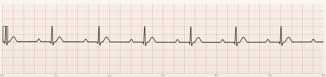
- P before every QRS, QRS after every P (1:1)
- PR longer than ~1 large box (>200 ms) and constant
- No dropped QRS complexes
How to say it: “Sinus rhythm with first-degree AV block” (name the base rhythm + the delay).
2. Second-Degree Type I — Mobitz I (Wenckebach)
Plain English: The PR keeps getting longer… longer… then a P shows up with no QRS. Then the PR resets short and the pattern often repeats (grouped beating).
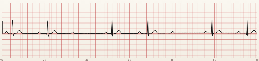
- Some P waves without a QRS (dropped beats)
- PR progressively longer on conducted beats
- Often “groups” of beats separated by a pause
Memory hook: Walking PR → then a miss. If the PR is walking, it is not Mobitz II.
3. Second-Degree Type II — Mobitz II
Plain English: Conducted beats share the same PR. Then — without warning — a QRS is missing. No progressive lengthening.
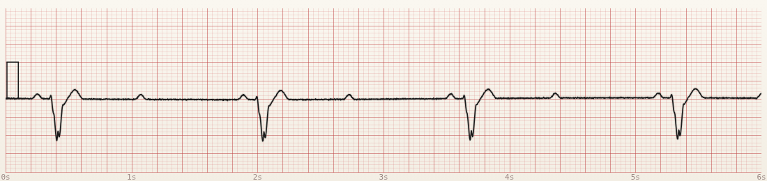
- Dropped QRS after some P waves
- PR the same whenever a beat does conduct
- QRS may be wide (classic teaching) — still use the PR pattern as the main differentiator from Mobitz I
Memory hook: Fixed PR → sudden miss. Educationally treated as higher risk for progression than classic Wenckebach — match care to the patient and protocol.
Note on 2:1 block: When every other P conducts, you may not see PR “walking.” Label carefully and use clinical context / protocol pathways rather than forcing a guess from one strip alone.
4. Third-Degree (Complete) Heart Block
Plain English: The atria fire on their own clock. The ventricles fire on a slow backup clock. Nothing is linking them — P waves march through the QRS and T waves.
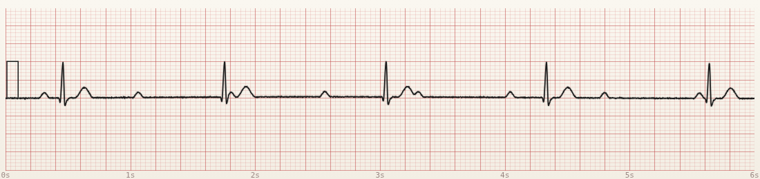
- Regular P–P and (usually) regular R–R — but unrelated to each other
- PR intervals that look random (because there is no real conduction)
- Escape QRS may be narrow (junctional) or wide (ventricular)
Memory hook: Two clocks, zero relationship. Not “sometimes drops” — never a consistent conducted pair.
Common Mix-Ups (Keep It Simple)
- Slow sinus vs Wenckebach: rate can change in sinus arrhythmia — Wenckebach needs a lengthening PR and a dropped QRS.
- Mobitz I vs II: walk = I. Fixed PR + drop = II. Do not skip measuring a few PRs in a row.
- Complete block vs “lots of P waves”: map several cycles. In 3°, P waves keep marching right through the ventricular complexes.
- Strip label vs patient: bradycardia + block still needs pulse, perfusion, and protocol — not only a correct name.
Decision Checkpoint
P waves are regular. Some P waves have no QRS after them. On the beats that do conduct, the PR looks identical each time — then a QRS disappears without the PR getting longer.
What is it? Mobitz II (fixed PR + sudden drop).
What next clinically? Assess the patient (pulse/perfusion/symptoms) and follow your bradycardia / block pathway and medical direction — the label is step one, not the whole plan.
Practice
Open CardioStrip and pull up each block again. For every strip say out loud:
- Every P a QRS?
- PR walking or fixed?
- One clock or two?
Pair with Systematic ECG Approach so you never skip P waves and PR under stress.
Sources & Further Study
- Strip images generated with CardioStrip (Code 3 Workshop educational EKG simulator).
- Standard paramedic / ACLS teaching frameworks for atrioventricular blocks.
- Your local protocol and medical direction remain authoritative for care.
Education only. Apply learning within your protocols and medical direction. This is not clinical care guidance. Strips are simulated teaching examples, not patient recordings.